Test Name
PTT Incubated Mixing Study (PTTIM)
CPT Codes
85730
85610
85732
85670
85520
85390
Methodology
Clot Detection
Turnaround Time
1 – 3 days
Specimen Requirements
Volume:
4 mL
Minimum Volume:
1.5 mL
Specimen Type:
Plasma
Collection Container:
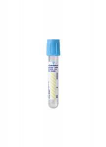
Light Blue Sodium Citrate Coagulation Tube
Transport Temperature:
Frozen

Specimen Collection & Handling
Discontinue coumadin therapy for 14 days prior to collection.
Discontinue direct thrombin inhibitors and heparin 2 days prior to collection.
Stability
Ambient:
7 days
Reference Range
See Interpretation
PT Screen:
8.4-13.0 seconds
APTT Screen:
< 33.2 seconds
APTT Immediate Mix:
33.2 seconds
APTT Incubated Mix:
< 36.0 seconds
Thrombin Time:
< 18.6 seconds
Heparin Assay:
< 0.1 U/mL
Additional Information
Background Information
The activated partial thromboplastin time (APTT) is one of the most commonly used tests to investigate bleeding patients, monitor anticoagulant therapy, and screen patients before surgery. The APTT measures the integrity of the intrinsic and common pathways of the coagulation cascade. The prothrombin time (PT), another common screening test, measures the integrity of the extrinsic and common coagulation pathway.
The APTT is measured as the number of seconds for the patient’s plasma to form a fibrin clot after the addition of an intrinsic pathway activator, phospholipid, and calcium. A prolonged APTT can be caused by a coagulation factor deficiency or the presence of an inhibitor. The mixing study, incubated APTT, is used to investigate the cause of a prolonged APTT result. The mixing study is performed by measuring the APTT in the patient’s plasma, then mixing an equal volume of the patient’s plasma and normal pooled plasma (NPP), and repeating the APTT tests immediately and after one-hour incubation.
The components of the panel include PT screen, APTT screen, APTT Immediate Mix, and APTT Incubated Mix, as well as a thrombin time and heparin anti-Xa assay if needed.
The principle of the mixing study can be summarized as:
1. If the prolonged APTT screen is due to a factor deficiency, mixing with an equal volume of NPP (which has approximately 100% of all coagulation factors) will replace the patient’s deficient factor. This result in an APTT immediate mix is shortened or corrected into the reference range.
2. If the prolonged APTT screen is due to the presence of an inhibitor, mixing with an equal volume of NPP will not shorten or correct the prolongation of APTT in repeated tests. The reason is the inhibitor in the patient’s plasma is present in excess and binds to coagulation factors or protein/phospholipid complexes in both the patient’s plasma and NPP.
Correction of the APTT in the mixing study suggests a coagulation factor deficiency in either the intrinsic pathway (factors VIII, IX, XI and XII, high-molecular-weight kininogen [HMWK] or prekallikrein [PK]), or in the common pathway (also prolonged PT) such as factor II, V, and X. Deficiency of factors VIII, IX, and XI will present with bleeding; however, deficiency of factor XII or prekallikrein will not increase bleeding risk, but may increase thrombotic risk. Further testing, such as clotting factor assays, is necessary to diagnose a specific factor deficiency. See Figure 1 for the diagnostic algorithm used in the laboratory.
There are three different types of inhibitors:
1. Inhibitors directly against specific factors, such as factor VIII or factor V inhibitors.
2. Anticoagulants such as heparins, fondaparinux, dabigatran, and other direct thrombin inhibitors.
3. Non-specific inhibitors, such as lupus anticoagulants.
Some inhibitors will demonstrate a delayed-type inhibitor pattern, with time and/or temperature dependence. In cases with a delayed-type inhibitor, the APTT Immediate Mix will correct to within the reference range; however, the APTT Incubated Mix will be prolonged.
Although rare, the presence of a factor inhibitor, such as a factor VIII inhibitor, will increase the risk of life-threatening bleeding. The presence of a factor inhibitor can be confirmed by a Bethesda assay for that factor. The presence of heparins, fondaparinux, dabigatran, or other direct thrombin inhibitors can cause prolongation of both the APTT Immediate Mix and APTT Incubated Mix. Careful clinical and medication history, and additional thrombin time with heparin assay (anti-Xa inhibition assay) can exclude the presence of anticoagulants.
The presence of lupus anticoagulants, which are antibodies against protein-phospholipid complexes, will increase the risk of thromboembolism. The presence of low-level, nonspecific inhibitors in the patient’s plasma may demonstrate a prolonged APTT Incubated Mix similar to a delayed-type inhibitor. If the clinical history suggests a lupus anticoagulant, further testing, including phospholipid based screening tests, phospholipid dependency assays, and exclusion of the presence of inhibitors, in addition to mixing study, incubated APTT, is necessary (refer to Figure 1 for lupus anticoagulant).
The adequate performance of the mixing test and accurate interpretation is important because the presence of a specific factor inhibitor, non-specific inhibitors, such as lupus anticoagulant, anticoagulants, or factor deficiency, have different clinical manifestations and require different clinical management.
Clinical Indications
The mixing test, incubated APTT, is indicated when the cause of a prolonged APTT result needs to be investigated.
Interpretation
APTT Screen:
Prolonged
APTT, Immediate Mix:
Normal
APTT, Incubated Mix:
Normal
If the APTT Screen is prolonged with a normal APTT Immediate Mix and APTT Incubated mix, this indicates a factor deficiency in the intrinsic or final common pathway.
If the PT is normal, this suggests an intrinsic pathway deficiency (VIII, IX, XI, XII, PK, HMWK).
If the PT is prolonged, this suggests a common pathway deficiency (fibrinogen, II, V, X).
APTT Screen:
Prolonged
APTT, Immediate Mix:
Normal
APTT, Incubated Mix:
Abnormal
If the APTT Screen is prolonged, with a normal APTT Immediate Mix, but an abnormal APTT Incubated Mix, this indicates the presence of a delayed inhibitor such as specific factor inhibitors, most commonly factor VIII inhibitor, and small numbers of lupus anticoagulant.
APTT Screen:
Prolonged
APTT, Immediate Mix:
Abnormal
APTT, Incubated Mix:
Abnormal
If the APTT Screen is prolonged, with an abnormal APTT Immediate Mix and abnormal APTT Incubated Mix, this favors a non-specific inhibitor, such as a lupus anticoagulant, and anticoagulants such as heparin, fondaparinux, dabigatran, or other direct thrombin inhibitors.
Methodology
The PT Screen is performed using Innovin® (Dade Behring, Inc.) reagent and STAR Evolution® Analyzer (Diagnostica Stago, Inc.). The PT Screen is included to localize abnormalities to common, intrinsic, and extrinsic pathways.
The APTT Screen is performed using the PTT-Automate reagent and STAR Evolution® analyzer (both Diagnostica Stago, Inc).
The mixing studies are performed by mixing the patient’s plasma with an equal volume of the NPP (Cryocheck; Precision Biologic, Inc).
For the APTT Immediate Mix, the APTT is performed immediately after mixing the plasmas. For the APTT Incubated Mix, the APTT is performed after one-hour incubation at 37ºC. The NPP serves as a negative control; two levels of positive control are performed; lupus positive plasma (Precision Biologic, Inc) and weak lupus positive plasma (Precision Biologic, Inc).
The thrombin time (Diagnostica Stago, Inc) will be measured in specimens with prolonged APTT. If the TT is prolonged, a heparin assay (anti-Xa inhibition assay; Rotachrom Heparin kit, Diagnostica Stago, Inc.) by a chromogenic assay will be performed to distinguish a heparin effect from a direct thrombin inhibitor.
Suggested Reading
1. Kottke-Marchant K. An Algorithmic Approach to Hemostasis Testing. CAP Press (2008).
2. Devreese KMJ. Interpretation of normal plasma mixing studies in the laboratory diagnosis of lupus anticoagulants. Thrombosis Research, 2007;119(3):369-376.
3. Favaloro EJ, Bonar R, Duncan E, Earl G, Low J et al. Misidentification of factor inhibitors by diagnostic haemostasis laboratories recognition of pitfalls and elucidation of strategies. A follow up to a large multi-center evaluation. Pathology. 2007;39(5):504-511.
4. Kamal AH, Tefferi A, Pruthi RK. How to interpret and pursue an abnormal prothormbin time, activated partial thromboplastin time, and bleeding time in adults. Mayo Clinic Proceedings. 2007;82(7):864-873.
