D-Dimer to Rule Out Venous Thromboembolism

Technical Brief:

D-Dimer to Rule Out Venous Thromboembolism


Test Name

D-Dimer (DDMER)

CPT Codes

85379

Methodology

Turbidimetric Immunoassay (TUI)

Turnaround Time

1 – 8 hours

Specimen Requirements

Volume:
2 mL

Minimum Volume:
1 mL

Specimen Type:
Plasma

Collection Container:
Light Blue Sodium Citrate Coagulation Tube

Transport Temperature:
Frozen
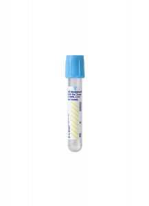
3.2% sodium citrate is the preferred anticoagulant recommended by CLSI.

Reference Range

< 500 ng/mL FEU

Clinical Information

Useful in the evaluation of DIC and fibrinolytic abnormalities.

Useful in the evaluation of deep vein thrombosis.

A negative result (< 500 ng/mL) may be helpful in the exclusion of venous thrombosis.

Additional Information

Background Information

Venous thromboembolism is a serious medical problem that can escalate rapidly to a life-threatening situation in the form of pulmonary embolism. The annual incidence of pulmonary embolism in the United States is estimated at 100,000 to 300,000 cases.1

Symptoms of venous thromboembolism or deep vein thrombosis (DVT) include pain and swelling in the affected arm or leg and associated erythema, tenderness, and warmth in the affected limb and calf pain on foot dorsiflexion. Symptoms typically are unilateral and include dyspnea, pleuritic chest pain, hemoptysis, low-grade fever, and tachycardia.

The D-dimer has proven most useful in patients suspected of having a pulmonary embolism and who have a low pretest probability of disease. Use of the Wells Criteria has shown to be a reliable and reproducible means of determining this pretest probability. While DVT cannot be completely ruled out or confirmed with the Wells Criteria, it can help inform the interpretation of subsequent diagnostic tests and reduce the need for invasive testing.

An estimated 70 percent of patients presenting in the emergency room with symptoms of DVT do not have the disorder.1 Therefore, a means for rapidly and accurately differentiating between patients with DVT and those without is critical to defining subsequent appropriate therapy to prevent pulmonary embolism.

Several techniques have been proposed in the past decade for detecting small, deep thrombi, including CT venography, duplex scanning, and MRI. These techniques, while effective, are time-consuming, expensive, and, in the case of CT venography, expose patients to radiation.2

The D-dimer assay offers a rapid, non-invasive, relatively inexpensive in vitro method to rule out venous thromboembolism, DVT, or pulmonary embolism. The assay provides a quantitative measure of D-dimer.

  • For suspected DVT, a D-dimer level below 500 ng/mL FEU has a Negative Predictive Value (NPV) of 99.2% and a sensitivity of 98.9 %.
  • For suspected pulmonary embolism, a D-dimer below 500 ng/mL FEU has an NPV of 99.1% and a sensitivity of 97.8%.

The initial insult to the vein initiates the coagulation cascade. Fibrin produced during this process, together with platelet, forms the thrombus at the site of injury. During subsequent fibrinolysis, plasmin cleaves factor XIIIa–crosslinked fibrin into several intermediate forms, including D-dimer fragments. D-dimer fragments, resulting from terminal fibrin degradation, are composed of smaller segments of crosslinked fibrin and are produced only during fibrinolysis.

Under normal conditions, D-dimer is undetectable in the blood. Therefore, D-dimer is a marker for intravascular coagulation, and its presence in the blood is suggestive of thrombosis.

To make a definitive diagnosis of venous thromboembolism, DVT, or pulmonary embolism, patients with an elevated D-dimer should undergo additional testing such as ultrasonography, ventilation-perfusion lung scan, and chest computed tomography (CT).3

Clinical Indications

Testing rules out venous thromboembolism, DVT, or pulmonary embolism in the presence of risk factors and clinical symptoms.

Interpretation

Negative: D-dimer < 500 ng/ml

Limitations

D-dimer should not be used as a stand-alone test.

Elevated D-dimer may be due to recent surgery, trauma, or infection.

Elevated levels are also seen with liver disease, pregnancy, eclampsia, cardiovascular disease, and some cancers.

Suggested Reading

1. Bockenstedt P. D-Dimer in Venous Thromboembolism. N Engl J Med. 2003;349:1203-1204.

2. Stein PD, Matta F. Acute pulmonary embolism. Curr Probl Cardiol. 2010 Jul;35(7):314-76.

3. Salaun PY, Couturaud F, LE Duc-Pennec A, Lacut K, LE Roux PY, Guillo P, Pennec PY, Cornily JC, Leroyer C, LE Gal G. Non-invasive diagnosis of pulmonary embolism. Chest. 2010; Aug.19, e-pub ahead of print.

Collagen Binding Activity Assay for von Willebrand Disease

Technical Brief:

Collagen Binding Activity Assay for von Willebrand Disease


Test Name

Colagen Binding Assay (CBA)

For further evaluation and classification, we suggest the von Willebrand Diagnostic Interpretive Panel (VWFPN).

CPT Codes

83520

Methodology

Enzyme-Linked Immunosorbent Assay (ELISA)

Turnaround Time

1 – 3 days

Specimen Requirements

Volume:
2 mL

Specimen Type:
Plasma

Collection Container:
Light Blue Sodium Citrate Coagulation Tube

Transport Temperature:
Frozen

3.2% sodium citrate is the preferred anticoagulant recommended by CLSI.

Specimens other than 3.2% trisodium citrate plasma are unacceptable.

Specimen Collection & Transport

Collection of blood by routine venipuncture in a 3.5 mL light blue top tube containing a 9:1 ratio of blood to 3.2% trisodium citrate anticoagulant.

Pediatric volume of 2.5 mL with an appropriate ratio of anticoagulant is acceptable.

Reference Range

CBA:
41-161%

Ratio of CBA/VWF:
Ag >=0.73

Background Information

Von Willebrand disease (VWD) is the most common inherited bleeding disorder with a prevalence of approximately 1% in the general population. It also can occur as an acquired bleeding disorder. VWD is a clinically heterogeneous disorder with several subtypes due to deficiency and/or dysfunction of von Willebrand factor (VWF). VWF is a multimeric adhesive glycoprotein that plays a major role in primary hemostasis and coagulation. VWF mediates adhesion of platelets to injured subendothelium and to the platelet surface receptor GPIb and serves as the specific carrier protein for coagulation factor VIII (fVIII) in plasma preventing proteolytic degradation. The revised classification of VWD identified two major categories: quantitative and qualitative defects.

The quantitative VWF defects include type 1 (partial deficiency of VWF) and type 3 (complete absence of VWF) in plasma and/or platelets. Type 2 is a qualitative VWF defect that is further classified as four subtypes by different pathophysiologic mechanisms.

Accurate laboratory diagnosis and classification of VWD using both quantitative (antigenic) and qualitative (functional) assays based on the VWD diagnostic algorithm are crucial because the presenting biological activity of VWF determines both the hemorrhagic risk and subsequent clinical management.

Abbreviations:

Ag: Antigen
CBA: Collagen-binding activity
fVIIl:C: Factor VIII: Coagulant
MW: Molecular weight

NL: Normal
PFA-100: Platelet function screen
PT: Prothrombin time
PTT: Partial thromboplastin time

RiCof: Ristocetin cofactor
RIPA: Ristocetin-induced platelet aggregation
VWF: von Willebrand factor

Clinical Indications

The functional activity of VWF traditionally has been assessed using the ristocetin cofactor activity (RiCof) assay, which measures the VWF-mediated agglutination of platelets in the presence of ristocetin. However, the usefulness of this assay has limitations due to poor reproducibility and lack of calibration. The collagen-binding activity (CBA) assay has been proposed as a supplemental test for VWF activity.

The CBA assay is based on the ability of multimeric forms of VWF to bind collagen, and its greatest strength lies in the ability to selectively detect primarily high molecular weight (HMW) forms of VWF, which are known to be most functional and adhesive.

The CBA assay is a useful adjunctive to the RiCof assay for the diagnosis of VWD and to differentiate VWD with deficiency of HMW multimer forms in type 2A and type 2B from type 1. It also can differentiate very low levels of VWF in severe type 1 from a complete absence of VWF in type 3 and has been reported as a better marker for therapeutic efficacy of treatment with DDAVP® (desmopressin) and fVIII concentrate.

Interpretation

CBA results are reported as % of the reference value for CBA. CBA to VWF:Ag ratio is calculated to provide a ratio of VWF activity to protein amount.

1. Type 1 VWD patients have concordantly decreased CBA and VWF:Ag levels.

2. Type 3 VWD patients have markedly decreased or nearly absent CBA and VWF:Ag levels.

3. Type 2A VWD and type 2B VWD patients have discordantly decreased CBA and VWF:Ag levels with markedly decreased CBA level, normal, or decreased VWF:Ag level and loss of HMW multimers.

4. Type 2M VWD patients have a discordantly decreased CBA level with a normal or decreased VWF:Ag level but without the loss of HMW multimers.

5. Type 2N VWD patients have normal CBA and VWF:Ag with discordantly decreased FVIII coagulant activity.

6. CBA values are known to be lower in O blood groups compared with non-O blood groups. However, as VWF:Ag levels show similar blood group dependence, the ratio of CBA/VWF:Ag is not affected.

Methodology

The CBA assay is an enzyme immunoassay (REAADS® Collagen Binding Assay ELISA kit, Corgenix, Inc., Broomfield, Colo.) that quantitates the binding of VWF to a collagen-coated microwell plate. After binding peroxidase-conjugated anti-VWF antibodies to VWF multimers, the resulting color intensity is determined photometrically, which is proportional to HMW forms of VWF present in the plasma. In situ evaluation for precision and accuracy of the CBA assay shows low coefficient of variation (6.3-11.1%) with a lower limit of detection 0.2% (linearity 1-530%).

Suggested Reading

1. Favaloro EJ. Von Willebrand factor collagen-binding (activity) assay in the diagnosis of von Willebrand disease: a 15-year journey. Seminar Thromb Hemost. 2002;28:191-202.

2. Nichols WL et al. The diagnosis, evaluation, and management of von Willebrand disease. U.S. Department of Health and Human Services. NIH Publication No. 08-5832, December 2007.

3. Castaman G, Federici AB, Rodeghiero F, Mannucci PM. Von Willebrand disease in the year 2003: toward the complete identification of gene defects for correct diagnosis and treatment. Haematologica. 2003;Jan;88(1):94-108.

4. Kalla A, Talpsep T. The von Willebrand factor collagen binding activity assay: clinical application. An Hematol. 2001;80:466-471.

5. Favaloro EJ. Toward a new paradigm for the identification and functional characterization of Von Willebrand disease. Seminar Thromb Hemost. 2009;35:60-75.

High-Sensitivity Flow Cytometry for Paroxysmal Nocturnal Hemoglobinuria

Technical Brief

High-Sensitivity Flow Cytometry for Paroxysmal Nocturnal Hemoglobinuria


Test Name

PNH Panel by FCM (PNHPNL)

CPT Codes

88184
88185 (x2)
88187

Methodology

Flow Cytometry (FC)

Turnaround Time

1 – 3 days

Specimen Requirements

Type:
Whole blood

Volume:
4 mL

Minimum Volume:
2 mL

Tube/Container:
Lavender BD Hemogard™ K2EDTA Tube

Transport Temperature:
Ambient

Clearly indicate specimen source on sample label.

Stability:

Plasma

Ambient: 
48 hours

Refrigerated:
Unacceptable

Frozen:
Unacceptable

Reference Ranges

Negative:
No PNH clone detected

Background Information

Paroxysmal nocturnal hemoglobinuria (PNH) is a clonal stem cell disorder characterized by mutations in the PIGA gene leading to loss of cell surface proteins linked to glycosylphosphatidylinositol (GPI) anchors. Patients affected by PNH display complement-mediated hemolysis, thrombosis, and bone marrow failure, though the clinical presentation is variable. The presence of a PNH clone occurs in classical hemolytic PNH, generally at levels above 1%; however, PNH clones may also be seen in other disorders such as aplastic anemia and myelodysplastic syndrome. Flow cytometric immunophenotypic analysis is the method of choice to detect populations of GPI anchor-deficient cells used in the diagnosis of PNH and to monitor patients with an established diagnosis.1 PNH erythrocyte clones may be divided into those with partial loss of CD59 (PNH Type II Cells) or complete loss of CD59 (PNH Type III Cells).

Cleveland Clinic Laboratories offers high sensitivity flow cytometry testing for paroxysmal nocturnal hemoglobinuria using whole blood. This procedure labels red blood cells (RBC) and white blood cells (WBC) for the detection of GPI-linked surface antigens using monoclonal antibodies and a fluorochrome-labeled bacterial aerolysin (FLAER).2 Through high sensitivity flow cytometry testing, as few as 0.01% PNH cells can be detected.1

Clinical Indications

This test may be useful in the evaluation of patients with intravasuclar hemolysis, unexplained hemolysis, thrombosis with unusual features, or bone marrow failure.

Interpretation

Results are reported as:

  • Negative
  • Low-level PNH clone positive (.01 – <1%) with percentage
  • PNH clone positive (≥1%) with percentage

Limitations

Results of PNH flow cytometry studies must be interpreted in the context of the clinical, laboratory, and histologic findings.

Blood not collected in a K2EDTA tube (or one which has been improperly stored and handled prior to receipt) cannot be processed.

Blood stability limit for PNH testing is 24 hours after the stated draw time. Clinical significance of results on specimens 24-48 hours old should be evaluated in the context of other clinical and laboratory findings.

Blood older than 48 hours from draw time cannot be analyzed.

Methodology

High sensitivity flow cytometry for PNH is performed in accordance with Clinical Cytometry Society recommendations for high sensitivity flow cytometry testing.1

Cells are stained directly with FITC, PE, PE/Cy7, PerCP/Cy5.5, APC, and APC/Cy7-labeled monoclonal antibodies to detect antigens of interest. Antibodies used include CD15, CD24, CD33, CD45, CD59, and glycophorin A. Additionally, FLAER is employed.

For erythrocytes, antibodies to glycophorin A are used to specifically gate red cells, and PNH clones are identified by lack of CD59 expression. PNH erythrocyte clones are divided into those with partial loss of CD59 (PNH Type II Cells) or complete loss of CD59 (PNH Type III Cells).

For granulocytes, CD15 and CD33 are used to specifically gate granulocytes. PNH-type granulocytes are then identified by lack of expression of CD24 and lack of reactivity to FLAER.

References

1. Borowitz MJ, Craig FE, DiGiuseppe JA et al. Guidelines for the Diagnosis and Monitoring of Paroxysmal Nocturnal Hemoglobinuria and Related Disorders by Flow Cytometry. Cytometry B Clin Cytom. 2010; 78B:211-230.

2. Sutherland DR, Kuek N, Davidson J et al. Diagnosing PNH with FLAER and Multiparameter Flow Cytometry. Cytometry B Clin Cytom. 2007; 72B: 167-177.

M. tuberculosis Complex versus Non-Tuberculous Mycobacteria by Polymerase Chain Reaction (PCR) on Smear-Positive Specimens

Technical Brief:

M. tuberculosis Complex versus Non-Tuberculous Mycobacteria by Polymerase Chain Reaction (PCR) on Smear-Positive Specimens


Test Name

MTB Complex vs NTM by PCR on Smear Positive Specimens (TBPCR)

CPT Codes

87551

Methodology

Polymerase Chain Reaction (PCR)

Turnaround Time

7 days

Specimen Requirements

Specimen Type:
Smear-positive bronchoalveolar lavage (BAL)

Volume:
5 mL

Minimum Volume:
1.5 mL

Collection Container:
Sterile specimen container

Transport Temperature:
Frozen

If aliquoting is necessary, sterile aliquot tubes must be used.

Specimen Type:
Smear-positive sputum

Volume:
1 mL

Collection Container:
Sterile specimen container

Transport Temperature:
Frozen

If aliquoting is necessary, sterile aliquot tubes must be used.

Specimen Type:
Smear-positive pleural fluid

Volume:
5 mL

Collection Container:
Sterile specimen container

Transport Temperature:
Frozen

If aliquoting is necessary, sterile aliquot tubes must be used.

Specimens for mycobacteria should be decontaminated and digested prior to freezing or long-term storage.

Freeze at -70°C and ship overnight.

Alternative Specimen

Specimen Type:
Smear-positive tissue

Volume:
3 cubic mm

Collection Container:
Sterile specimen container

Transport Temperature:
Ambient

3 cubic mm fresh undigested tissue should be frozen at -70 Celsius and shipped overnight.

Stability 

Ambient: 
Unacceptable

Refrigerated: 
Unacceptable

Frozen: 
Resp. – 1 year if frozen within 72 hours

Tissue – 2 years if frozen within 24 hours

Background Information

Mycobacterium tuberculosis infects one-third of the world’s population and is the leading cause of death due to any infectious agent worldwide. The incidence of non-tuberculous mycobacteria (NTM) infections is increasing, and NTM isolates now are more common in the United States than M. tuberculosis. Strict isolation is required under the Centers for Disease Control and Prevention guidelines for all patients suspected of having tuberculosis; isolation is not required for patients infected with NTM. Treatment of tuberculosis and NTM also differs.

The ability to rapidly and accurately distinguish M. tuberculosis from NTM has significant clinical implications. This information should dictate appropriate infection control measures and guide the selection of appropriate antimicrobial therapy. The LightCycler system (Roche Diagnostics, Indianapolis, Ind.) combines real-time PCR with fluorogenic hybridization probes using fluorescence resonance energy transfer probes. This assay achieves rapid PCR results and has high sensitivity and specificity for the majority of clinically relevant mycobacteria, including M. tuberculosis, when smear-positive specimens are tested. Melting curve analysis performed by the LightCycler allows for differentiation of M. tuberculosis from NTM.

Clinical Indications

Detection and differentiation of M. tuberculosis from NTM on smear-positive specimens.

Culture for Mycobacterium spp. should be performed on all specimens ordered for acid-fast bacilli because of the possibility of dual mycobacterial infections and to have the isolate available for susceptibility testing if appropriate.

Interpretation

Results are reported qualitatively as positive or negative for M. tuberculosis, and positive or negative for NTM.

Limitations

This assay, as well as commercially-available assays, is insensitive when smear-negative specimens are tested.

This assay has suboptimal sensitivity for some of the rapidly growing Mycobacterium species and M. xenopi.

Methodology

The LightCycler FastStart DNA Master Hybridization Probe Kit (Roche) is used in conjunction with broad-range mycobacterial PCR primers and specially designed FRET hybridization
probes. Rapid-cycle PCR and post-amplification melt curve analysis are performed in the LightCycler system.

References

1. Shrestha NK, Tuohy MJ, Hall GS, Reischl U, Gordon SM, Procop GW. Detection and Differentiation of Mycobacterium tuberculosis and nontuberculous mycobacterial isolates by real-time PCR. J Clin Microbiol. 2003;41:5121-6.

Fecal Occult Blood Tests for Colorectal Cancer Screening

Technical Brief:

Fecal Occult Blood Tests for Colorectal Cancer Screening


Test Name

Fecal Occult Blood Test (IFOBT)

CPT Codes

82274

Methodology

Immunoassay (IA)

Turnaround Time

4 days

Specimen Requirements

Specimen Type:
Stool

Collection Container:
OC-Auto® FIT Personal Use Kit

Transport Temperature:
Ambient

Bulk stool (stool not contained in a collection vial) will be rejected.

Record date and time of collection on the test vial in permanent marker – do not use pencil or ballpoint pen.

Patients should be instructed to place the inoculated test vial and a copy of the test order into the pre-addressed mailing envelope.

Stability 

Ambient: 
15 days

Refrigerated: 
30 days at 4°C

Frozen: 
Unacceptable

Reference Range

Negative for hemoglobin at < 100 ng/ml

Background Information

In the United States, colorectal cancer is the third most common cancer diagnosed among men and women, and the second leading cause of death from cancer. Colorectal cancer can largely be prevented by the detection and removal of adenomatous polyps. Five-year survival is 90% if the disease is diagnosed while still localized, but only 68% for regional disease (a disease with lymph node involvement), and only 10% if distant metastases are present.1 In spite of this, a majority of U.S. adults are not receiving regular age- and risk-appropriate screening or have never been screened at all.2

There is a range of options for colorectal cancer screening that includes stool testing for the presence of occult blood or exfoliated DNA, or structural examinations that include flexible sigmoidoscopy, colonoscopy, double-contrast barium enema, and CR-colonography. Beginning in 1980, the American Cancer Society first issued formal guidelines for colorectal cancer screening in average-risk individuals and these have been periodically updated, along with additional guidelines for high-risk individuals.3 Other organizations, such as the American College of Radiology, U.S. Preventive Services Task Force, and the U.S. Multi-Society Task Force on Colorectal Cancer, have also issued recommendations. Collaborative efforts among these groups took place in 2008 to come to a consensus and provide joint guidelines and recommendations.1

Occult gastrointestinal bleeding refers to bleeding that is not apparent to the patient. It has traditionally been identified by tests that detect fecal blood, or, if bleeding is sufficient, as iron deficiency anemia. A variety of fecal occult-blood (FOB) tests have been designed primarily to screen for colon cancer, including both guaiac-based (gFOB) and immunochemical-based stool tests (iFOBT or FIT). The likelihood that FOB tests will detect gastrointestinal blood is affected by the type of test utilized, anatomical level of bleeding (upper GI vs. colonic), stool transit time, stool mixing, intra-luminal hemoglobin degradation, and features intrinsic to bleeding lesions (i.e. irregular bleeding).4

Clinical Indications

There are two major reasons to detect occult blood in the stool: as a screen for colorectal cancer or to detect upper or lower GI bleeding. In the outpatient settings, an immunochemical-based assay (iFOBT or FIT) is used to detect fecal occult blood for colorectal cancer screenings; the three-part guaiac-based fecal occult blood test will no longer be available for screening.

  • The single guaiac card (SENSA) will continue to be used for the detection of bleeding (upper or lower GI bleeding).
  • The iFOBT (FIT) cannot be used for detection of upper GI bleeding and should only be used as a colon cancer screen test.
  • The HemaPrompt card (a guaiac-based POCT assay) was introduced in October 2009 for use as a point-of-care test for detection of upper and lower GI bleeding.
    • It can be used in the Emergency Department and a few other clinical areas that have been granted POCT testing privileges.

If colorectal cancer screening is the intended use of the occult blood test, the iFOBT is the preferred method because it is the more-sensitive screen, and only a single sample collection is required.

One of the main reasons for the change to iFOBT for colorectal cancer screening is that the guaiac-based tests lack the sensitivity and specificity seen with the iFOBT assays. In an early study of iFOBT assays, specimens obtained from 107 colorectal cancer subjects showed that the iFOB test had a 97% sensitivity for detection of colorectal cancer, as compared to a very-sensitive guaiac-based test (Hemoccult Sensa) in which the sensitivity was 94%. The iFOBT also demonstrated greater sensitivity (76%) in detecting large adenomas as compared with guaiac-based tests, in which the sensitivity was 42%. The specificity of the iFOBT was 97.8%, as compared to 96.1% for the gFOB, when 1,355 screening tests were performed. The authors concluded that the iFOBT provided the best combination of specificity and sensitivity.5 In a more recent study of performance characteristics for detecting cancer, the sensitivity of the iFOBT test was 81.8% vs. 64.3% with gFOB test.

Specificity for iFOBT was 96.9% and 97.3%, respectively, for detecting cancer and adenomas versus 90% and 90.6% with gFOB. For the detection of large polyps, the sensitivity of the gFOB was actually higher than that with iFOBT in this study.6 In a recent study out of Seoul, Korea, a population of average-risk individuals (770 patients from four centers) undergoing colorectal cancer screening were assayed for occult blood comparing a gFOB to an iFOB. The iFOBT provided a higher sensitivity for detecting cancer and advanced colorectal neoplasia than the gFOB and had an acceptable specificity that could significantly reduce the need for colonoscopic evaluation of the screened population.7 An editorial in the same issue of the journal from Seoul suggested that the data supported an effort to increase the use of iFOBT assays in the U.S. and publishing of studies in average-risk individuals here as well.8

Secondly, various foods and exogenous substances can yield false-positive guaiac-based FOB test results. Vitamin C in excess of 250 mg/day from all sources (dietary and supplemental) can oxidize guaiac. False-positive guaiac-based test results can also occur with excess dietary red meat, such as beef, lamb, processed meat, liver, or plant peroxidases contained in raw fruits and vegetables, especially radishes, turnips, horseradish, cantaloupes, and other melons. Such foods should be avoided for 72 hours prior to testing. GI bleeding can be induced by alcohol, and also has well-known iatrogenic causes, such as steroids and NSAIDS.9

The iFOBT assays detect human globin, a protein that along with heme constitutes human hemoglobin. It is more specific for human blood as compared to the guaiac-based tests, and is not subject to the false-negative results seen in the guaiac-based tests in the presence of high dose vitamin C supplements. It is important to remember that the iFOBT assays are specific to bleeding in the lower GI tract.

Thirdly, because of the increased sensitivity of these assays, only one sample is usually required for screening, as compared to the three samples needed when guaiac-based tests are used. The ease of use of these screening assays should enhance patient compliance.

Interpretation

Positive:
Samples with a hemoglobin concentration >/=100 ng/mL

Negative:
Samples with a hemoglobin concentration <100 ng/mL

Because gastrointestinal lesions may bleed intermittently, and blood in feces is not distributed uniformly, a negative result may not assure the absence of a lesion.

Limitations

Patients with the following conditions should not be tested due to a potential for false positive results:

  • Bleeding hemorrhoids
  • Menstrual bleeding
  • Constipation bleeding
  • Urinary bleeding

Certain medications, such as aspirin and non-steroidal anti-inflammatory drugs, may cause gastrointestinal irritation and subsequent bleeding in some patients, which may cause false positive results.

Contamination of the sample with urine or toilet water also may cause erroneous results.

The OC-Sensor Diana iFOBT should not be used for testing urine, gastric or other body fluids.

Because iFOB (FIT) tests are dependent on the intact antigenic structure of heme molecules, its use is limited to screening for FOB that is not gastric or upper GI in origin.

Bulk stool samples should not be sent to the lab for occult blood testing, as hemoglobin present in stool begins to degrade within hours of passage.

Methodology

The OC-Sensor Diana iFOB test is an immunoassay-based test that uses rabbit polyclonal antibodies to detect hemoglobin in stool. The test is a turbidimetric latex agglutination test. Hemoglobin present in the patient sample will combine with latex-coated antibody to cause a change in absorbance. A light beam is passed through the reaction cells and measures changes in the intensity of light beam. Testing is performed on an automated analyzer and qualitative results are generated.

Patients should be provided with a collection kit, which is composed of a labeled collection vial, instructions on how to collect the stool sample and inoculate the vial, and an envelope into which the vial can mailed to the laboratories for testing.

Testing is performed on a daily basis, Monday through Friday

References

1. Levin B, Lieberman DA, McFarland B et al. Screening and Surveillance for the early detection of colorectal cancer and adenomatous polyps, 2008: a joint guideline from the American Cancer Society, the U.S. Multi-Society Task Force on Colorectal Cancer, and the American College of Radiology. Ca Cancer J Clin. 2008;58:130-60.

2. Smith RA, Cokkinides V, Eyre HJ. Cancer screening in the U.S., 2007: a review of current guidelines, practices, and prospects. CA Cancer J Clin. 2007;57:90-104.

3. Rex DK et al. American College of Gastroenterology Guidelines for Colorectal Cancer Screening 2008. The Am J Gastroenterol. 2009;104:739-50.

4. Rockey, DC. Occult gastrointestinal bleeding. N Engl J Med. 1999;341:38-46.

5. St. John DJ, Young GP, Alexeyeff MA et al. Evaluation of new occult blood tests for detection of colorectal neoplasia. Gastroenterology. 1993;104:1861-8.

6. Sakoda LC, Levin TR et al. Screening for colorectal neoplasms with new fecal occult blood tests: update on performance characteristics. J Natl Cancer Institute. 2007;99:1462-70.

7. Park D, Ryu S, Kim YH et al. Comparison of guaiac-based quantitative immunochemical fecal occult blood testing in a population at average risk undergoing colorectal cancer screening. Am J Gastroenterol. 2010;105:2017-25.

8. Allison JE. Editorial: FIT: a valuable but underutilized screening test for colorectal cancer – it’s time to change. Am J Gastroenterol. 2010;105:1026-8.

9. Bakerman, S. ABC’s of interpretive laboratory data. Fourth Edition. Interpretive Laboratory Data Inc. Scottsdale, AZ. 2002.

BK Polyomavirus by Real-Time PCR (Detection & Quantitation)

Technical Brief:

BK Polyomavirus by Real-Time PCR (Detection & Quantitation)


Test Name

BK Virus Quantitation PCR, Plasma (BKQUAN)

CPT Codes

87799

Methodology

Polymerase Chain Reaction (PCR)

Turnaround Time

5 days

Specimen Requirements

Volume:
2 mL

Minimum Volume:
1 mL

Specimen Type:
Plasma

Collection Container:
Lavender BD Hemogard™ K2EDTA Tube

Transport Temperature:
Refrigerated

Plasma must be separated within 24 hours of collection.  If aliquoting is necessary, sterile aliquot tubes must be used.

Stability 

Ambient: 
Unacceptable

Refrigerated: 
5 days

Frozen: 
30 days

Clinical Information

Reportable range is from 500 – 5,000,000 copies/mL

Reference Range

Negative for BKV DNA

Background Information

Acquired in childhood or adolescence, the polyomavirus (BKV) is one of a few polyomavirus infections that infect humans. The virus is spread from person to person and is most likely transmitted through the respiratory pathway.

Although its prevalence is high — 60 to 90% of people have antibodies to the BK virus — the infection usually remains latent, and individuals rarely demonstrate signs and symptoms. However, the virus is important in immunocompromised hosts, where reactivation of the latent infection causes disease.

This virus has emerged as an important cause of renal allograft infection (i.e. BK nephropathy). The BK virus also is associated with urethral stenosis, interstitial nephritis, and is one of the causes of hemorrhagic cystitis in bone marrow transplant recipients.1

The use of immunosuppressive drugs, which are necessary to prevent immunologic rejection of the renal allograft, has the side effect of increasing the likelihood of opportunistic infections. This immunosuppressed state affords the reactivation of a latent BK virus; and, if viral replication remains unchecked, then BK nephropathy will develop. It is estimated reactivation of the BKV occurs in 10 to 60% of renal transplant recipients, with anywhere from 1 to 5% developing BK nephropathy.2,3

Screening and early intervention for BKV have been a major advance over the past six to seven years and has led to an approximately eight-fold reduction in graft loss due to BKV. Most kidney transplant centers now employ BKV screening using quantitative BKV tests.

The presence of the BK virus may be assessed through a qualitative PCR of the urine. If the BK virus is present, then the quantity of the BK polyomavirus present is important. The higher the viral load in blood, the more likely the presence of renal disease. A progressive rise in serum creatinine concentration in a kidney transplant recipient, regardless of the underlying cause, should prompt a referral for reevaluation by the transplant center.

Treatment of BK nephropathy remains poorly defined. Most often, a decrease in immunosuppression is the therapy of choice, but this must be carefully balanced with the increased risk of renal allograft rejection.4

Clinical Indications

The BK virus most commonly produces asymptomatic infections; however, for patients with kidney and bone marrow transplants, BK virus infections are a cause of morbidity and mortality. Therefore, the quantitative BKV by PCR assay is indicated for renal transplant patients who are known to harbor the BK virus, particularly those with deterioration in renal function.

The assay is only to be used in patients with appropriate risk factors for BK-associated disease and is not indicated for the screening of asymptomatic patients.

International consensus guidelines have established the blood viral load of 10,000 copies/ml as a common threshold for intervention.5 In most patients, the quantity of the BK virus can be reduced with prompt restoration of BK virus-specific immunity, frequent monitoring, and timely modification or reduction of immunosuppression. According to an international consensus panel (Hirsch HH, 2005), monitoring for BK virus is recommended every three months for the first two years post-renal transplant or when allograft dysfunction occurs.

Methodology

Quantitative real-time PCR for the BK virus in the blood is currently the only noninvasive test for the measurement and monitoring of the BK viral load, which is important for the diagnosis and assessment of the treatment of BK nephropathy.

Interpretation

Results are reported in copies/mL of polyoma (BKV) virus. Detection of BKV DNA in clinical specimens supports the clinical diagnosis of renal disease due to BKV. The presence of BKV DNA in plasma at levels ≥ 10,000 copies BKV DNA/mL is specific for polyomavirus-associated nephropathy (PVAN).

Limitations

A negative result does not rule out the possibility of BK virus (BKV) infection; clinical correlation is necessary. Repeat testing at an appropriate interval may be needed.

References

1. WE Braun: BK polyomavirus: A newly recognized threat to transplanted kidneys. Cleveland Clinic Journal of Medicine. Dec 2003;1056-1068

2. Ramos E, Drachenberg CB, Papadimitrion JC, et al. Clinical course of polyoma virus nephropathy in 67 renal transplant patients. J Am Soc Nephrol. 2002;13:2145-2151.

3. Shah KV. Human polyomavirus BKV and renal disease. Nephrol DialTransplant. 2000;15:754-755.

4. S. Hariharn, Kidney International. 2006;69:655-662.

5. Hirsch HH, Brennan DC, Drachenberg CB, et al. Polyomavirus-associated nephropathy in renal transplantation: interdisciplinary analyses and recommendations. Transplantation. May 2005;79(10):1277-86